


It’s safer and more effective.
Amanda Bradbury’s first signs of epilepsy began with visual auras when she was 19. As time passed, she experienced frequent anxiety, struggled to focus, had trouble following conversations, and even faced difficulty speaking and swallowing.
“I would get an overwhelming sense of fear before a seizure, which I now know was seizure-related,” Bradbury, now 29, shared in a news release. “I started isolating myself because I was nervous about how seizures affected my memory. Talking became a challenge, and I often felt confused. I became more unsure of what was happening around me.”
Bradbury was diagnosed with focal epilepsy, where seizures stem from a misfiring area in the brain. In her case, the problem was in her amygdala, the brain’s emotion center, explaining the fear she felt before and during seizures.
Doctors eventually cured her epilepsy through surgery that removed the lesion causing the seizures after medications failed to control them. Now, a breakthrough in medical imaging may allow more epilepsy patients to have similar outcomes.
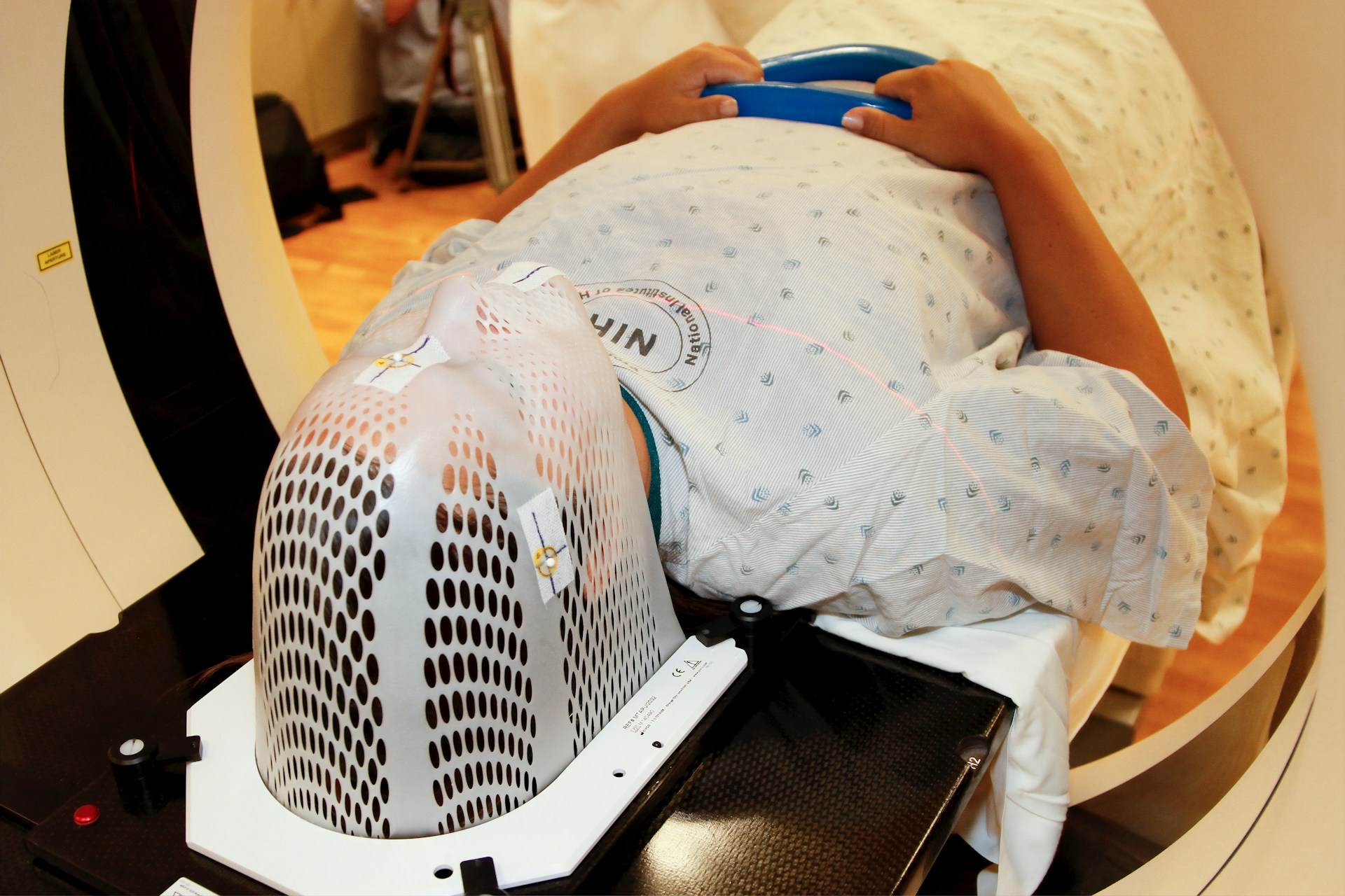
Bradbury’s brain lesion was large enough to be detected by standard MRI scanners. However, many epilepsy patients have lesions that are too small to be seen with conventional imaging. Researchers have discovered that 7 Tesla (7T) MRI scanners, which create stronger magnetic fields than standard ones, can now identify small, previously undetectable epilepsy-causing lesions deep in the brain.
Historically, 7T MRI scanners struggled with signal blackspots in key brain areas, especially in the temporal lobes, where most epilepsy cases originate. To address this, researchers employed a technique called “parallel transmit,” using eight transmitters around the brain instead of just one.
“Much like how a single Wi-Fi router leaves areas with weak signals, older MRI scanners left blackspots on brain scans,” explained Chris Rodgers, a professor of biomedical imaging at the University of Cambridge. “By using multiple transmitters around the head, we get clearer images with fewer blackspots, which is crucial for pinpointing the brain area causing seizures.”
The technique was tested on 31 epilepsy patients who didn’t respond to medication. The enhanced 7T scanner revealed previously unseen lesions in nine patients and confirmed suspected lesions in four others. In some cases, the scan even disproved the suspected causes of the seizures. The advanced imaging changed epilepsy management for 18 of the 31 patients: nine were offered surgery to remove the lesions, while others received alternative treatments.
“Epilepsy that doesn’t respond to medication can greatly impact a person’s life, affecting their independence and ability to work,” said Dr. Thomas Cope, a neurology lecturer at the University of Cambridge. “We can cure many of these patients, but only if we can accurately locate the source of their seizures. This new scanning technique will make life-changing surgery accessible to more patients.”
Before her surgery, Bradbury’s brainwave tests revealed she was having multiple seizures a day, far more than the few she believed were occurring weekly. Post-surgery, she noticed significant improvements, feeling more energized, less anxious, and better able to focus.
“Even during recovery, it was clear the surgery was the right choice,” Bradbury said. “I suddenly realized I could do so much more. I started wondering what else I could accomplish.”
From simple tasks like cleaning the kitchen to pursuing bigger dreams, Bradbury’s life has transformed. Although she had to leave an interior design course due to her epilepsy, she now works in office administration and plans to reignite her passion for interior design as a hobby.
“I want to enjoy things like interior design and creative activities again,” she said. “Now, I feel like I can experience so much more.”


Discussion about this post